Blog
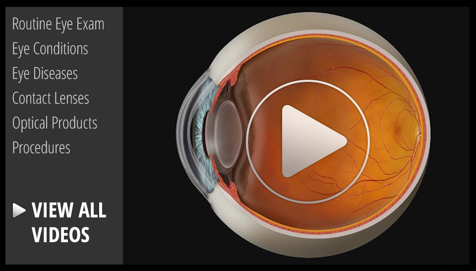
The retina is the nerve tissue that lines the inside back wall of your eye. Light travels through the pupil and lens and is focused on the retina, where it is converted into a neural impulse and transmitted to the brain. If there is a break in the retina, fluid can track underneath the retina and separate it from the eye wall. Depending on the location and degree of retinal detachment, there can be very serious vision loss.
Symptoms
The three 3 F’s are the most common symptoms of a retinal detachment:
Flashes: Flashing lights that are usually seen in peripheral (side) vision.
Floaters: Hundreds of dark spots that persist in the center of vision.
Field cut: Curtain or shadow that usually starts in peripheral vision that may move to involve the center of vision.
Causes
Retinal detachments can be broadly divided into three categories depending on the cause of the detachment:
Rhegmatogenous retinal detachments: Rhegmatogenous means “arising from a...
Read more: What's a retinal detachment and what are the symptoms?

We sometimes get asked, "Why do I need an eye exam when I can see great?"
An eye exam doesn't just check your visual acuity--we are also looking for a number of treatable eye diseases that have few or no visual symptoms in their early stages. In fact, the three leading causes of legal blindness in the United States all start with almost no visual symptoms detectable by the person wit the disease. The three diseases are macular degeneration, glaucoma, and diabetic retinopathy. Each of these diseases gets more prevalent as people age. That is why regular eye exams are recommended to become more frequent as adults get older.
Macular Degeneration: The leading cause of legal blindness in the United States is a treatable--but not curable--disease. Early detection and treatment can significantly improve the long-term outcome. In the earliest stages, often when people are unaware that they have a problem, treating the disease with a very specific vitamin regimen called AREDS 2 can...


