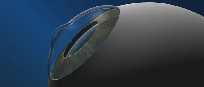
Keratoconus is an eye disease in which the cornea deforms from its normally curved dome shaped and becomes cone shaped. Sometimes there is a flaw in the collagen, the material of the cornea that weakens and allows the cornea to stretch into an irregular cone shape.
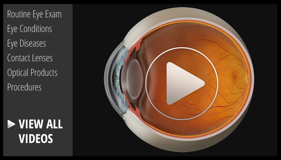
The cornea is the clear tissue located at the front of the eye and it refracts and focuses light as it enters the eye. Therefore abnormalities of the corneal surfaces can severely distort vision.
Symptoms usually start in the teen years with near-sighteness and astigmatism which can often be treated with contact lenses or glasses. At the onset it can be difficult to detect.
It is first diagnosed when the cornea starts reveal progressive irregular distortion and eventually becomes to advance for conventional glasses or contact lenses to correct. At this critical point it is often treated with specially designed contact lenses to provide a smooth optical surface to focus light rays and impede progression of the bulging cone shape.
In some case keratoconus can progress to the point that corneal replacement surgery is needed. This usually occurs around the age of 35.
There are some evidence to suggest allergy suffers and people with rigid contact lenses that rub their eyes may contribute to the progression of the symptoms and cause scratches on the surface of the cornea.
If you feel you are at risk or your prescription is changing rapidly make certain your eye care professional checks for Keratoconus during your next eye health and visual examination.